DESCRIPTION
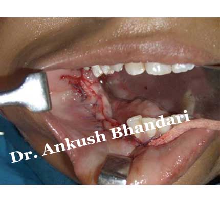
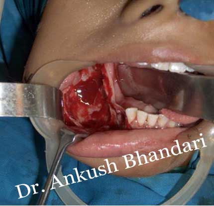
- Enucleation — Removal of the entire cyst
- Marsupialization — The creation of a window into the wall of a cyst, allowing the contents to be drained. The window is left open, and the lack of pressure within the cyst causes the lesion to shrink, as the surrounding bone starts to fill in again.
- Enucleation following marsupialization—sometimes marsupialization is carried out as a single procedure, but usually it is followed by a second procedure to remove the cyst. This is particularly the case when cysts are very large and their removal would leave a significant surgical defect.
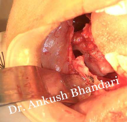
- Enucleation with curettage—this is the removal of the cyst and some of the surrounding bone, which may contain some of the lining of the cyst.
Diagnosis :-
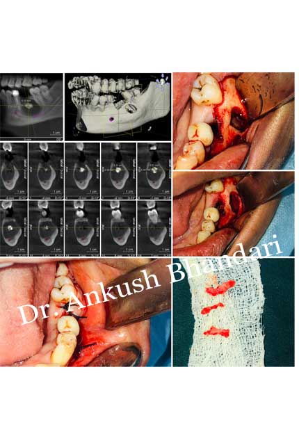
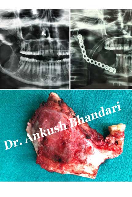
Most cysts are discovered as a chance finding on routine dental radiography. On an x-ray, cysts appear as radiolucent (dark) areas with radiopaque (white) borders. Cysts are usually unilocular, but may also be multilocular. Sometimes aspiration is used to aid diagnosis of a cystic lesion; e.g., fluid aspirated from a radicular cyst may appear straw-colored and display shimmering due to cholesterol content. Almost always, the cyst lining is sent to a pathologist for histopathologic examination after it has been surgically removed. This means that the exact diagnosis of the type of cyst is often made in retrospect.
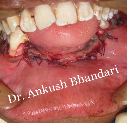
Treatment:-
When treatment is required, this is usually by surgical removal of the cyst. There are four ways in which cysts are managed.